
- Information
- AI Chat
Chapter 3 workbook Cells and Tissues
Human Anatomy and Physiology I (BIO 203 )
Hagerstown Community College

Comments
- Bravo it helped me and my colleagues alot
Preview text
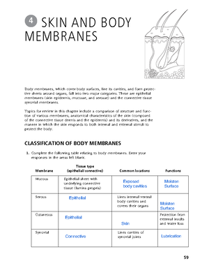
The basic unit of structure and function in the human body is the cell. Each of a cell’s parts, or organelles, as well as the entire cell, is organized to perform a specific function. Cells have the ability to metabolize, grow and reproduce, move, and respond to stimuli. The cells of the body differ in shape, size, and in specific roles in the body. Cells that are similar in structure and function form tissues, which, in turn, construct the various body organs.
Student activities in this chapter include questions relating to the structure and function of the generalized animal cell and to the general arrangement of tissues and their contribution to the activities of the various body organs.
CELLS
Overview
- Answer the following questions by inserting your responses in the answer blanks. _________________________ 1. _________________________ 2. _________________________ 3. _________________________ 4. _________________________ 5. _________________________ 6. _________________________ 7. _________________________ 8. _________________________ 9. _________________________ 10.
33
CELLS AND
TISSUES
3
1–4. Name the four elements that make up the bulk of living matter. 5. Name the single most abundant molecule in living matter. 6. Name the trace element most important for making bones hard. 7. Name the element, found in small amounts in the body, that is needed to make hemoglobin for oxygen transport. 8–12. Although there are many specific “jobs” that certain cells are able to do, name five functions common to all cells. _________________________ 11. _________________________ 12.
_________________________ 13.
_________________________ 14.
_________________________ 15.
_________________________ 16.
_________________________ 17.
Anatomy of a Generalized Cell
- Using the list of terms on the following page, correctly label all cell parts indicated by leader lines in Figure 3–1. Then, select different colors for each structure and use them to color the coding circles and the corre- sponding structures in the illustration.
34 Anatomy & Physiology Coloring Workbook
13–15. List three different cell shapes. 16. Name the fluid, similar to seawater, that surrounds and bathes all body cells. 17. Name the flattened cells, important in protection against damage, that fit together like tiles. (This is just one example of the generalization that a cell’s structure is very closely related to its function in the body.)
Cytosol Vacuole
Figure 3–
- Based on Figure 3–3, answer the following: (A) Label the specializations of the plasma membrane. (B) Color the coding circles and the corresponding cell parts. (C) Answer the questions provided below.
○ Nucleus ○ Nucleolus ○ Mitochondrion ○ ER
- What type of cell function(s) does the presence of microvilli typically indicate? __________________________________________________________________________________
- Which cell junction forms an impermeable barrier? _________________________________________
- Which cell junction is an anchoring junction? _______________________________________________
- Which junction has linker proteins spanning the intercellular space? ________________________
- Which cell junction is not illustrated, and what is its function? ______________________________
- Which two types of membrane junctions would you expect to find between cells of the heart? _______________________________________________ and ____________________________________________
36 Anatomy & Physiology Coloring Workbook
A
B
C
Figure 3–
- Relative to cellular organelles, circle the term or phrase that does not belong
in each of the following groupings. Then, fill in the answer blanks with the
correct group name.
- Peroxisomes Enzymatic breakdown Centrioles Lysosomes Group: _________
- Microtubules Intermediate filaments Microfilaments Cilia Group: _________
- Ribosomes Smooth ER Rough ER Amino acids Group: _________
- Double membrane Cristae ATP production Vitamin A storage Group: _________
- Centrioles Mitochondria Cilia Flagella Group: _________
- ER Ribosomes Transport vesicles Golgi apparatus Group: ________
- Nuclear pores DNA Lysosomes Chromatin Nucleolus Group: _________
- Name the cytoskeletal element (microtubules, microfilaments, or intermediate filaments) described by each of the following phrases. _________________________ 1. Give the cell its shape _________________________ 2. Resist tension placed on a cell _________________________ 3. Radiate from the cell center _________________________ 4. Involved in moving intracellular structures _________________________ 5. Are the most stable _________________________ 6. Have the thickest diameter
- Different organelles are abundant in different cell types. Match the cell types with their abundant organelles by selecting a letter or letters from the key choices. Items may have more than one answer.
Key Choices
A. Golgi apparatus C. Lysosomes E. Mitochondria G. Rough ER B. Intermediate filaments D. Microfilaments F. Peroxisomes H. Smooth ER ______ 1. Cell lining the small intestine (assembles fats) ______ 2. White blood cell; a phagocyte ______ 3. Liver cell that detoxifies carcinogens ______ 4. Muscle cell (contractile cell) ______ 5. Mucus-secreting cell (secretes a protein product) ______ 6. Cell at external skin surface (withstands friction and tension) ______ 7. Kidney tubule cell (makes and uses large amounts of ATP)
Chapter 3 Cells and Tissues 37
Chapter 3 Cells and Tissues 39
ABC
Figure 3–
- Figure 3–6 is a simplified diagram of the plasma membrane. Structure A represents channel proteins constructing a pore, structure B represents an ATP- energized solute pump, and structure C is a transport protein that does not depend on energy from ATP. (A) Identify these structures and the membrane phospholipids by color.
○ Channel ○ Solute pump ○ Passive transport protein carrier ○ Phospholipids
(B) For each substance that moves through the plasma membrane, draw an arrow indicating its (most likely) direction of movement (into or out of the cell). If it is moved actively, use a red arrow; if it is moved passively, use a blue arrow. Color the coding arrows. Active Passive
Figure 3–
Cell exterior
Cell interior
Steroid
Amino acid
Amino acid
Fat H 2 O CO 2
CO 2
O 2
O 2
Cl–
Na+
Na+
Glucose
Glucose
K+
K+
(C) Answer the following questions referring to Figure 3–6:
- Which of the substances shown moves passively through the lipid part of the membrane? ______________________________________________________________________
- Which of the substances shown enters the cell by attachment to a passive transport protein carrier? _______________________________________________________________
- Which of the substances shown moves passively through the membrane by moving through its pores? __________________________________________________________
- Which of the substances shown would have to use a solute pump to be transported through the membrane? ___________________________________________________
- Select the key choices that characterize each of the following statements. Insert the appropriate letter(s) or corresponding term(s) in the answer blanks. Items may have more than one answer.
Key Choices
A. Active transport D. Exocytosis G. Phagocytosis B. Diffusion, simple E. Facilitated diffusion H. Pinocytosis C. Diffusion, osmosis F. Filtration I. Receptor-mediated endocytosis _________________________ 1. Engulfment processes that require ATP _________________________ 2. Driven by concentration gradient _________________________ 3. Driven by hydrostatic (fluid) pressure (typically blood pressure in the body) _________________________ 4. Moves down a concentration gradient _________________________ 5. Moves up (against) a concentration gradient; requires a carrier _________________________ 6. Moves small or lipid-soluble solutes through the membrane _________________________ 7. Transports amino acids and Na+ through the plasma membrane _________________________ 8. Examples of vesicular transport _________________________ 9. A means of bringing fairly large particles into the cell _________________________ 10. Used to eject wastes and to secrete cell products _________________________ 11. Membrane transport using channels or carrier proteins that does not require ATP
40 Anatomy & Physiology Coloring Workbook
- Identify the phases of mitosis depicted in Figure 3– 7 by inserting the correct name in the blank under the appropriate diagram. Then, select different colors to represent the structures listed below and use them to color in the coding circles and the corresponding structures in the illustration.
○ Nuclear membrane(s), if present ○ Centrioles
○ Nucleoli, if present ○ Spindle fibers
○ Chromosomes
42 Anatomy & Physiology Coloring Workbook
A
C
B
D Figure 3–
- The following statements describe events that occur during the different phases of mitosis. Identify the phase by choosing the correct response(s) from the key choices and inserting the letter(s) or term(s) in the answer blanks. Items may have more than one answer.
Key Choices
A. Anaphase C. Prophase E. None of these B. Metaphase D. Telophase _________________________ 1. Chromatin coils and condenses to form deeply staining bodies. _________________________ 2. Centromeres break, and chromosomes begin migration toward opposite poles of the cell. _________________________ 3. The nuclear membrane and nucleoli reappear. _________________________ 4. When chromosomes cease their poleward movement, this phase begins. _________________________ 5. Chromosomes align on the equator of the spindle. _________________________ 6. The nucleoli and nuclear membrane disappear. _________________________ 7. The spindle forms through the migration of the centrioles. _________________________ 8. Chromosomal material replicates. _________________________ 9. Chromosomes first appear to be duplex structures. _________________________ 10. Chromosomes attach to the spindle fibers. _________________________ 11. A cleavage furrow forms during this phase. _________________________ 12. The nuclear membrane is absent during the entire phase. _________________________ 13. A cell carries out its usual metabolic activities.
- Using the key choices, complete the crossword puzzle by answering each of the clues provided.
Key Choices
Anucleate Centromeres Cytoplasm Nucleus Aster Centrosomes Interphase Prophase Binucleate Coiled Loose Spindle Across 3. The structure that acts as a scaffolding for chromosomal attachment and movement is called the _______. 4. If a cell undergoes nuclear division but not cytoplasmic division, the product is a _________ cell. 7. Chromosomes attach to the spindle fibers by undivided structures called ______.
Chapter 3 Cells and Tissues 43
- Transfer of the genetic message from DNA to mRNA is called _________________________.
- Assembly of amino acids according to the genetic information carried by mRNA is called _________________________.
- The set of three nitrogen bases on tRNA that is complementary to an mRNA codon is called a _________________________. The complementary three-base sequence on DNA is called a _________________________.
Chapter 3 Cells and Tissues 45
Nucleus Nuclearmembrane
Ribosome
1 2
A
G C G A A C T T A T A
S am e str and
Figure 3–
46 Anatomy & Physiology Coloring Workbook
BODY TISSUES
- The four major tissue types are named in Figure 3–9. For each tissue type, provide its major function(s) on the lines after the tissue name. Then, list the location of each tissue type at the end of each leader line.
Nervous tissue:
Muscle tissue:
- Epithelial tissue:
Connective tissue:
Figure 3–
48 Anatomy & Physiology Coloring Workbook
- Describe briefly how the particular structure of a neuron relates to its function
in the body. _________________________________________________________________
G
I
H
J
KL Figure 3–10, G–L
- Using the key choices, correctly identify the major tissue types described. Enter the appropriate letter or tissue type term in the answer blanks.
Key Choices
A. Connective B. Epithelium C. Muscle D. Nervous _________________________ 1. Forms mucous, serous, and epidermal membranes _________________________ 2. Allows for organ movements within the body _________________________ 3. Transmits electrochemical impulses _________________________ 4. Supports body organs _________________________ 5. Cells of this tissue may absorb and/or secrete substances _________________________ 6. Basis of the major controlling system of the body _________________________ 7. Cells of this tissue shorten to exert force _________________________ 8. Forms hormones _________________________ 9. Packages and protects body organs _________________________ 10. Characterized by having large amounts of nonliving matrix _________________________ 11. Allows you to smile, grasp, swim, ski, and shoot an arrow _________________________ 12. Most widely distributed tissue type in the body _________________________ 13. Forms the brain and spinal cord
- Using the key choices, identify the following specific type(s) of epithelial tissue. Enter the appropriate letter or classification term in the answer blanks.
Key Choices
A. Pseudostratified columnar (ciliated) C. Simple cuboidal E. Stratified squamous B. Simple columnar D. Simple squamous F. Transitional _________________________ 1. Lines the esophagus and forms the skin epidermis _________________________ 2. Forms the lining of the stomach and small intestine _________________________ 3. Best suited for areas subjected to friction _________________________ 4. Lines much of the respiratory tract _________________________ 5. Propels substances (e., mucus) across its surface _________________________ 6. Found in the bladder lining; peculiar cells that slide over one another _________________________ 7. Forms thin serous membranes; a single layer of flattened cells
Chapter 3 Cells and Tissues 49
- Using the key choices, identify the following connective tissue types. Insert the appropriate letter or corresponding term in the answer blanks.
Key Choices
A. Adipose connective tissue C. Dense fibrous connective tissue E. Osseous tissue B. Areolar connective tissue D. Hyaline cartilage F. Reticular connective tissue _________________________ 1. Provides great strength through parallel bundles of collagenic fibers; found in tendons _________________________ 2. Acts as a storage depot for fat _________________________ 3. Composes the majority of the dermis of the skin _________________________ 4. Forms the bony skeleton _________________________ 5. Composes the lamina propria and packages organs; includes a gel-like matrix with all categories of fibers and many cell types _________________________ 6. Forms the embryonic skeleton and the surfaces of bones at the joints; reinforces the trachea _________________________ 7. Provides insulation for the body _________________________ 8. Matrix with no specific shape, heavily invaded with fibers; appears glassy and smooth _________________________ 9. Contains cells arranged concentrically around a nutrient canal; matrix is hard due to calcium salts _________________________ 10. Forms the stroma or internal “skeleton” of lymph nodes, the spleen, and other lymphoid organs
Tissue Repair
- For each of the following statements about tissue repair that is true, enter T in the answer blank. For each false statement, correct the underlined words by writing the correct words in the answer blank. _________________________ 1. The nonspecific response of the body to injury is called regeneration. _________________________ 2. Intact capillaries near an injury dilate, leaking plasma, blood cells, and antibodies, which cause the blood to clot. The clot at the surface dries to form a scab. _________________________ 3. During the first phase of tissue repair, capillary buds invade the clot, forming a delicate pink tissue called endodermal tissue. _________________________ 4. When damage is not too severe, the surface epithelium migrates beneath the dry scab and across the surface of the granulation tissue. This repair process is called proliferation.
Chapter 3 Cells and Tissues 51
_________________________ 5. If tissue damage is very severe, tissue repair is more likely to occur by fibrosis, or scarring. _________________________ 6. During fibrosis, fibroblasts in the granulation tissue lay down keratin fibers, which form a strong, compact, but inflexible mass. _________________________ 7. The repair of cardiac muscle and nervous tissue occurs mainly by fibrosis.
DEVELOPMENTAL ASPECTS OF CELLS AND TISSUES
- Correctly complete each statement by inserting your responses in the answer blanks. _________________________ 1. _________________________ 2. _________________________ 3. _________________________ 4. _________________________ 5. _________________________ 6. _________________________ 7. _________________________ 8. _________________________ 9. _________________________ 10. _________________________ 11. _________________________ 12. _________________________ 13. _________________________ 14. _________________________ 15. _________________________ 16. _________________________ 17. _________________________ 18. _________________________ 19. _________________________ 20.
52 Anatomy & Physiology Coloring Workbook
During embryonic development, cells specialize to form (1). Mitotic cell division is very important for overall body (2). All tissues except (3) tissue continue to undergo cell division until the end of adolescence. After this time, (4) tissue also becomes amitotic. When amitotic tissues are dam- aged, they are replaced by (5) tissue, which does not func- tion in the same way as the original tissue. This is a serious problem when heart cells are damaged. Aging begins almost as soon as we are born. Three explana- tions of the aging process have been offered. One states that (6) insults, such as the presence of toxic substances in the blood, are important. Another theory states that external (7) factors, such as X-rays, help to cause aging. A third theory suggests that aging is programmed in our (8). Three examples of aging processes seen in all people are (9) , (10) , and (11). Neoplasms occur when cells “go wild” and the normal con- trols of cell (12) are lost. The two types of neoplasms are (13) and (14). The (15) type tends to stay localized and have a capsule. The (16) type is likely to invade other body tissues and spread to other (distant) parts of the body. To cor- rectly diagnose the type of neoplasm, a microscopic examina- tion of the tissue called a (17) is usually done. Whenever possible, (18) is the treatment of choice for neoplasms. An overgrowth of tissue that is not considered to be a neo- plasm is referred to as (19). Conversely, a decrease in the size of an organ or tissue, resulting from loss of normal stimu- lation, is called (20).
Chapter 3 workbook Cells and Tissues
Course: Human Anatomy and Physiology I (BIO 203 )
University: Hagerstown Community College


- Discover more from:


