
- Information
- AI Chat
Head Trauma and TBI
Health And Illness I (NUR 275)
Pennsylvania College of Health Sciences
Recommended for you


Preview text
Head Trauma & TBI
Head Injury o Head injury: any damage to the head as a result of trauma o Brain injury: an external force that can cause significant damage to the brain o Head injury does NOT always mean brain injury o Traumatic brain injury (TBI): injury that is the result of an external force Causes & Risk Factors o Causes: Falls Motor vehicle accidents (MVA’s) Sport and recreation Assaults o Risk Factors: Age Infants & children 0- 4 yrs- physical abuse, shaken baby syndrome Adolescents 15-19 yrs Adults >65 years Men> women o Elderly highest risk= falls; teens highest risk= MVA’s Prevention o Prevention is the gold standard o Examples: Airbags Passive seat belts
Not drinking and driving Preventing falls at home Not speeding Not texting and driving Pathophysiology o TBI leads to swelling or bleeding increasing ICP o Increased ICP leaves no room for expansion in the cranium allowing for more increased ICP o Pressure on the blood vessels decreases perfusion to the brain o Cerebral hypoxia and ischemia occur o ICP continues to increase leading to herniation o Cerebral blood flow decreases Traumatic Injury
Primary Secondary Direct contact to the head/brain during the instant of initial injury
Occurs over hours to days as a result of the primary injury Focal: localized to one area of the brain
Inadequate delivery of nutrients and oxygen to the cells Diffuse: global injury to the brain
Injury Types o Scalp injuries Very vascular which can result in a lot of bleeding “Looks worse than it actually is” Stop the bleed, clean up the wound, suture it up High infection risk o Open head trauma: scalp laceration/tear in the dura, skull is opened (ex: bullet or ice pick)
Ex: baseball bat hits the occipital lobe the contrecoup will be seen in the frontal lobe Skull Fractures o Skull fracture: break in the continuity of the skull from forceful trauma o Linear: single fracture or crack in the bone Normally let it heal on its own and treatment is pain management o Comminuted: splintered/shattered Treatment is pain control and possible surgery o Depressed: bone fragments that become inwardly depressed into the brain tissue Treatment is pain control and surgery o S/S: pain at the site, swelling, warmth/bruising/redness, facial weakness o ** An ongoing headache indicates there is a fracture** Basal Skull Fracture o Basal skull fracture: fracture of the base of the skull o S/S: Facial trauma CSF leak from the ears or nose Blood leaking from the ears/nose Halo sign: take a sample of the draining fluid and put it on a white surface and will get
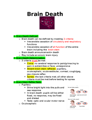
the halo sign; testing the CSF for glucose can help also determine if it is CSF Battle sign: bruising behind the ear Racoon eyes o Most occurs in the temporal lobe o NOTHING goes up the nose can cause intracranial tubing or placing the tube in the brain o *** Anytime you see CSF/blood from the ears/nose or racoon eyes or battle sign automatically think basilar*** o Assessment: Halo sign testing; yellow ring= + CSF Glucose test strip (+) = CSF No nasal suctioning Check nuchal rigidity (c-spine cleared 1st) Battles sign/racoon eyes o Treatment: High risk for infection so watch for s/s of infection Follow up with diagnostic testing Possible need for antibiotics High risk for bacterial meningitis Contusion o Contusion: bruising of the surface of the brain o Type of focal lesion (acceleration-deceleration injuries, coup & contrecoup) o Causes: Head trauma /blunt trauma Falls MVA’s Violence/ penetrating wounds Sport injuries o Found in the frontal/temporal lobe o More serious than a concussion o S/S:
o Acute: * hours* Changes in LOC Pupillary signs Hemiparesis Coma Cushing’s Triad (increase BP, decreased HR/RR) o Subacute: 48 hours – 2 weeks o Chronic: 2 weeks – months Seen commonly in the elderly who are prone to brain atrophy (a normal part of the aging process) Can be mistaken for a stroke Most often overlooked/forgotten or blamed on being a part of the aging process Blood becomes thicker and darker coming to look like motor oil with its consistency as well S/S: Severe headache coming and going Alternating focal neurological signs Personality changes Mental deterioration Focal seizures Treatment: Clot evacuation Reversal of coagulopathies Burr holes Craniotomy Intracerebral Hemorrhage o Intracerebral hemorrhage: a blood clot deep in the middle of the brain that is hard to remove o Pressure from the clot may cause brain damage
o Can be caused by nontraumatic events such as an aneurysm rupture, systemic HTN, bleeding disorders, or complications from anticoagulant therapy o S/S: Neurological deficits HA Change in LOC Increased ICP Focal changes (“thunder clap”) o Treatment: surgery to relieve the pressure Concussion o Concussion: temporary loss of neurologic function with no apparent brain damage Most common brain injury o S/S: Brief loss of consciousness HA Dizziness N/V Confusion Drowsiness Irritability Giddiness Visual & gait disturbances Normal neuro exam o Injury to the frontal lobe can present as bizarre irrational behavior, temporal lobe produces temporary amnesia or disorientation o NEVER leave someone with a bad concussion alone as they can develop traumatic encephalopathy o CT or MRI is used to diagnosis a concussion as there is no structural brain damage o Anticipate: HA, difficulty concentrating, amenia
Immediate coma Global cerebral edema Unexplained neuro deficit Assessments o ABC’s o Details of the injury o History LOC, amnesia, GCS o GSC: Mild TBI= 13-15 with LOC up to 15 min Moderate TBI= 9-12 with LOC up to 6 hours May have difficulty with work, learning, or role function Severe TBI= 3-8 with LOC >6 hours Requires management of hemodynamic status and intracranial pressure o The lower the GCS= greater the injury Medica Management
Manage ICP Osmotic diuretics: Mannitol Calcium channel blockers: prevent vasospasm
Ventilatory support
Prevent seizures Keppra Anticonvulsa nts
Fluid & electrolyte balance
Nutritional support Within 24 hours needed to think of this
Decrease anxiety Anxiety + Pain= increase in ICP Pain control Avoid opioids due to change in LOC and effect it can
Evacuate clots Debridement
have on the neuro checks Elevate skull fractures
Craniectomy Burr holes
Bone flaps Ventriculostomy Allows for ICP monitoring and draining
Family support and gift of life as this can lead to brain death
Assessment & Nursing Problems
Respiratory Ineffective airway clearance Impaired gas exchange
Neurologic R/F ineffective cerebral tissue perfusion
Cardiovascular Decreased cardiac output
Integumentary R/F impaired skin integrity
Musculoskeletal Impaired mobility
GI
Pain-decreased motility GU Increased risk for infection, skin breakdown incontinence
Metabolic Imbalanced nutrition less than body requirements
Psychological Ineffective coping
Nursing Interventions o Maintain the airway Vents, O2, suction, prevent aspiration, ABG’s, oral care o Neuro assessments LOC, GCS, neuro checks, reflexes, pupils, ICP, CPP Prevent increasing ICP o Monitor VS, CM, end tidal Co2, temperature, CSF leaks
o Caregiver role strain
Head Trauma and TBI
Course: Health And Illness I (NUR 275)
University: Pennsylvania College of Health Sciences


- Discover more from:
