- Information
- AI Chat
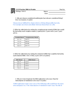
Bio 1107 Unit 3 Review
Principles Of Biology I (BIOL 1107)
University of Georgia
Recommended for you


Preview text
Biology 1107 Unit 3 Review
| Mitotic Division
Cell Cycle: cells that go through cell division go through regulated stages called the cell cycle which consists of two major phases: interphase and mitotic phase – then replicated DNA and cytoplasm separates into two daughter cells
Interphase: 3 phases - G1 (first gap): the cell performs normal biological functions but grows and accumulates materials and energy needed to move on to S phase - S (synthesis of DNA): DNA replication takes place, each chromosome consists of two chromatids firmly attached to each other at the centromere region - G2 (second gap): cell replenishes energy stores and makes final preparations for cell division Mitotic Phase (M phase): steps where the duplicated chromosomes line up, separate, and move into two new identical daughter cells – cell undergoes structural changes enabling it to move chromosomes around the cell and to maximize that chromosomes will be distributed equally - Mitosis (karyokinesis): chromosomes separate to form two nuclei o Prophase: chromosomes condense microtubules assemble and form the spindle o Prometaphase: nuclear envelope fragments and disperses microtubules of the spindle begin attaching to kinetochores on each chromatid o Metaphase: chromosomes fully condensed and move to middle of cell align on the metaphase plate between two poles of the cell chromatids are tightly attached to each other and face opposite sides of cell o Anaphase: Chromatids separate from each other at the centromere by the spindle and kinetochores to opposite ends of the cell Each chromatid is now called a chromosome o Telophase: Chromosomes reach opposite poles and begin to decondense Microtubule spindle disassembles and will reform to be part of cytoskeleton Separate nuclear envelopes form around two groups of chromosomes - Cytokinesis: cytoplasmic contents are divided to form two separate cells, differs between plant and animal cells o Animal cells (no cell walls) Ring of actin cytoskeleton fibers (contractile ring) forms at the site of the metaphase plate
Motor proteins cause fibers to slide relative to each other and pull the membrane in to form a cleavage furrow Actin fibers continue to slide past each other and the furrow deepens and splits the cell in two o Plant cells (cell wall) Rigid cell wall prevents formation of a cleavage furrow Vesicles containing phospholipids, protein, and cell wall components are transported to the site of the metaphase plate Fuse together to form a cell plate As more vesicles fuse the cell plate enlarges until it splits the cytoplasm into two separate cells
Regulation: generally, cells will only divide if the cell is ready to initiate division and if it receives signals from outside indicating that it is appropriate for the cell to divide - G1 checkpoint: determines if all conditions are good for cell division to proceed o Cell needs enough raw material, energy stored, and for the DNA to be in good condition (minimal/no damage), and cell receiving signals o If conditions no met cell won’t enter S phase and can attempt to fix the problem or wait for more signals from outside the cell o If checkpoint fails then the cell may divide to produce more cells than are needed or cells with damaged DNA Uncontrolled cell division = cancer - G2 checkpoint: checks to determine if all chromosomes have been replicated and that the replicated DNA is not damaged o If problem is detected then the cell cycle is halted until replication is complete or DNA is repaired o If damage cannot be fixed, the cell will trigger apoptosis (cell death) to prevent formation of cells that have damaged DNA - M checkpoint: checks to determine if the chromatids are correctly attached to the spindle microtubules o If unattached kinetochores are detected the cell will not proceed into anaphase until the kinetochores of each pair of chromatids are anchored to at least two spindle fibers from opposite end of the cell o If the checkpoint fails nondisjunction can occur Nondisjunction chromosomes are distributed incorrectly into daughter cells Nondisjunction occurs from M checkpoint failure (spindle not correctly attached)
Example of diploid in mitosis
Meiosis I: produces two cells with half as many chromosomes as the original cell, each daughter cell contains only one of each homologous pair even though the chromosomes that are present consist of two chromatids - Interphase o G1 (first gap): the cell performs normal biological functions but grows and accumulates materials and energy needed to move on to S phase o S (synthesis of DNA): DNA replication takes place, each chromosome consists of two chromatids firmly attached to each other at the centromere region o G2 (second gap): cell replenishes energy stores and makes final preparations for cell division - Prophase I: as nuclear envelope starts to break down during prophase I, homologous chromosomes line up side by side and are held together by a complex of proteins o Synapsis: tight pairing of chromosomes that aligns genes on the chromatids with each other o The X and Y chromosomes are not homologous but share a homologous region - Prometaphase: homologous pairs of chromosomes are held together at the chiasmata; microtubules attach to the fused kinetochores off the sister chromatids o Chromosomes continue to condense and the nuclear envelope completely disappears - Metaphase I: homologous chromosomes randomly assemble at the metaphase plate where they have been maneuvered into place by microtubules - Anaphase I: o Spindle microtubules pull the homologous chromosomes apart to opposite sides of the cell o Sister chromatids are attached at the centromere - Telophase I and Cytokinesis: o Sister chromatids arrive at the poles of the cell and begin to decondense o Nuclear envelope forms around each nucleus and the cytoplasm is divided by a cleavage furrow o Result is two haploid cells o Each cell contains one duplicated copy of each homologous chromosome pair Meiosis II: four daughter cells with half the starting number of chromosomes are produced - Chromosomes are not replicated again instead the chromosomes are moved to the middle of the cell and the chromatids are separated during anaphase II like mitosis - Each dividing cell has half as many chromosomes as a cell undergoing mitotic division in organisms o Four daughter cells with half the starting number of chromosomes are produced - Prophase II o Sister chromatids condense o New spindle begins to form o Nuclear envelope starts to fragment - Prometaphase II o Nuclear envelope disappears, and spindle fibers engage the individual kinetochores on the sister chromatids - Metaphase II
o Sister chromatids line up at the metaphase plate
- Anaphase II o Sister chromatids are pulled apart by the shortening of the kinetochore microtubules o Non-kinetochore microtubules lengthen the cell
- Telophase II and Cytokinesis o Chromosomes arrive at the poles of the cell and decondense o Nuclear envelopes surround the four nuclei o Cleavage furrows divide the two cells into four haploid cells Genetic Variation
- Independent assortment o Need at least two pairs o During metaphase I homologous chromosomes line up in the middle of the cell independently
- Recombination o When homologous chromosomes align during prophase of meiosis I it enables chromatids to exchange equivalent chromosomal segments by recombination/crossing over Leads to recombinant chromatids
- Fertilization o Mixing of genes from two parents
| Life Cycles
Introduction - In sexual reproduction offspring are unique but also similar to parents o In sexually reproducing species, no two individuals (with the exception of identical twins) have the same DNA and each member of the population possesses somewhat different characteristics o Advantageous in changing environment Diploid dominance - Most cells present during the life of an organism are diploid and the only haploid cells are the gametes - Fusion of gametes during fertilization produces the first diploid cell of a new individual (zygotes) - Zygote undergoes mitosis and cytokinesis to produce all the new cells of an individual - Only a fraction of the cells in an individual (germ cells) will be capable of undergoing meiosis to produce new gametes - Most animals including humans are diploid dominant Haploid dominance - The body of the organism is haploid - In sexual reproduction special haploid cells from two individuals are designated as + and – mating types to join to form a diploid zygote - Diploid zygote goes meiosis and cytokinesis to form four haploid cells called spores o spores are haploid and contain a new combination of genes from the two parents
Bio 1107 Unit 3 Review
Course: Principles Of Biology I (BIOL 1107)
University: University of Georgia