- Information
- AI Chat
BIOL3014 - Molecular Biology of the Cell
Molecular cell biology (BIOL3014)
University of Southampton

Preview text
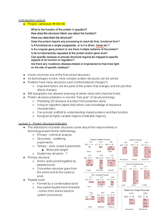
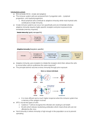
Co lour Key: Important Knowledge Needs Additional Reading to enhance Additional Reading Knowledge Needs research to explain further/don’t understand. Lecture 1 - An Overview of the Cell The discovery of cells is inextricably linked to the creation of the first microscopes Hooke’s microscope suffered from a poorly executed focusing mechanism and would tend to wear very quickly and unevenly. The lenses suffered from significant chromatic and spherical aberration but to correct this a small diaphragm was placed to reduce peripheral light rays and sharpen the image. Leeuwenhoek’s microscope was made with a ground lens not a blown lens and so solved the problem of chromatic/spherical aberration. It was so good it was able to discover microorganisms It wasn’t until the 19th century that universal cell theory came about. Light microscopy is limited by the wavelength of light so the best resolution possible is about 0. microns. Resolution could be extended by the visualisation of a moving object with fluorescence microscopy. EM microscopy has provided “near-atomic” resolution imaging and has vastly increased the depth of cell biology knowledge. Organelles were discovered through centrifugation. Fractionation Differential centrifugation Isopycnic centrifugation Lecture 2 - The Cytoskeleton Actin filaments are stained by rhodamine-labelled phalloidin whereas microtubules are labelled by primary antibodies which are recognised by secondary antibodies that are conjugated with fluorochromes. In the cellular cytoskeleton, 3 types of filaments: - Microtubules - Intermediate filaments - Actin filaments None of which can be resolved by conventional light microscopy. Epithelial cells are asymmetric (apical/basolateral), have a distinct shape and exist to transport things across themselves. They need a cytoskeleton. Actin comes in 3 forms: lamellipodia/filopodia and stress fibres.
Two alternate forms of actin machinery coexist at the leading edge of most motile cells. Lamellipodia which seem to be designed for persistent protrusion over a surface and filopodia which appear to perform sensory and exploratory functions to steer cells depending on cues from the environment In lamellipodia, branched nucleation of actin by the Arp2/3 complex, followed by filament elongation and barbed-end/(+)-end capping (which is necessary to keep filaments short and their number constant) In filopodia, both branching and capping need to be inhibited to allow continuous elongation of parallel filaments at their tip. They are initiated from a dendritic network by action of a tip complex that protects filaments from capping Lamellipodial organisation vs filopodial organisation is selected in part by the action of CP (capping protein) which acts as a negative regulator of filopodia formation. Stress fibres are bundles of actomyosin found in non-muscle cells that are the major mediators of cell contractions. They are composed of bundles of 10-30 actin filaments which are held together by actin-crosslinking protein 𝛼-actinin. Their structure is highly similar to that of the actomyosin arrays of the muscular sarcomere. Microtubules originate from the MTOC whereas actin filaments form from the nucleation of 3 G-actin subunits. Actin polymerisation arises from the formation of a complex of 3 actin monomers from which an actin monomer may elongate (a nucleus). Although a high concentration of G-actin exists in non-muscle cells, they alone fail to nucleate new microfilaments efficiently due to the instability of G-actin monomers and so need additional factors to nucleate. Tip nucleation involves formin proteins clustering at the plasma membrane and initiating the nucleation of actin filaments (whose structural integrity is maintained by fascin cross-linking). Convergent elongation involves Arp2/3 complex nucleated branches continually developing from the actin filament network located at the leading edge of the lamellipodia. These filaments gradually converge, forming a bundle that is secured by fascin cross-linking. Microtubule nucleation is typically spatially restricted to MTOCs. Temporal control is achieved by coupling the regulation of nucleation site assembly and/or activation to cell cycle profession or a specific point during cell differentiation. Microtubule polymerisation requires a nucleator which mimics or stabilises a small microtubule seed from multiple 𝛼𝛽-tubulin heterodimers. ℽ-tubulin localises to all known MTOCs and is required for the function, it is thought that the helical arrangement of ℽ-tubulin molecules in ℽTuRC matches the symmetry of a microtubule and so provides an assembly platform for 𝛼𝛽-tubulins through contacts between ℽ-tubulin and 𝛼-tubulin. Why do we need a cytoskeleton?
- To maintain cell structure (e. positioning of cell organelles)
- Maintaining cell shape and asymmetry (e. in neurons, epithelial cells and red blood cells)
- Maintaining cell polarity (e. the apical or basolateral ends of epithelial cells, in cell division)
- Allow for movement (intracellular transport, whole cell movement and amoeba) The cytoskeleton is highly dynamic but must also be stable - to achieve this all filaments are made of small protein subunits which are organised in polymers that are regulated tightly in space. Actin is the protein subunit which forms actin filaments. Each subunit has a globular 4 subunit structure and contains an ATP/ADP binding cleft. Actin is kept in an equilibrium between G- and F-actin so at lower concentration everything is in a globular form whereas at high concentrations,
The Arp2/3 complex is a large protein complex made up of 7 subunits which catalyse the formation of branched actin networks but requires the activation by a nucleation promoting factor to do this. Cell polarisation can be induced by the Rho GTPases (Rho, Rac and Cdc42) and by PIP signalling. The central function of RhoGTPases is to pass signals to effectors. → Rho proteins exist in the Rho-GDP bound form complexed with GDI (Guanine nucleotide Displacement Inhibitor), which retains them in the inactive state in the cytosol. Membrane bound signalling pathways bring Rho proteins to the membrane and through the action of a GEF, exchange the GDP for GTP, thus activating them. Membrane-bound activated Rho GTP can then bind effector proteins that cause changes in the actin cytoskeleton. Activation of Rho by lysophosphatidic acid (LPA) through an LPAR on the cell surface leads to either the formation of unbranched F-actin through a formin pathway or the activation of non-muscle myosin II by catalysing (through a Rho-activated kinase) the phosphorylation of the myosin light chain and the phosphorylation (and therefore inhibition) of the myosin light-chain phosphatase. Overall, this leads to a higher level of myosin light-chain phosphorylation and therefore contraction. So in the end, increased actin polymerisation and increased myosin activity leads to stress fibre formation and contraction. Rac activation is mediated by growth factors (e PDGF) and leads to lamellipodia formation via Arp2/3 through activation of the WAVE complex which leads to the assembly of branched actin filaments at the leading edge. Cdc42 activation leads to filopodia formation by stimulating actin assembly by Arp2/3 through activation of WASP. Twinfilin? Lecture 3: The Cytoskeleton and Movement F-actin has a 10 nm diameter where each repeat is 72 nm and is made of 14 subunits. F-actin has a right-handed double helical structure. Dissociation rates at both ends but association is higher at the (+)-end. Toxins are used to study actin filaments. Phalloidin stabilises filaments and so inhibits depolymerisation. Cytochalasin D and latrunculin promote depolymerisation. Auxiliary proteins are used to control actin polymerisation. Profillin binds to G-actin-ADP and catalyses ADP/ATP exchange and adds to (+) ends. Tropomodulin stabilises actin by capping the (-) end and inhibiting depolymerisation and so is used when stable structures are needed. Cofillin destabilises F-actin by by binding F-actin-ADP and leading to filament breaking which creates more pointed ends and so enhances net disassembly. Formin protects from CapZ binding and also brings 2 G-actin subunits together (nucleation) which can bind to profilin-actin-ATP complexes and so can grow the filament
The Arp2/3 complex creates branched networks. Intermediate filaments are intermediate in their size and lie between actin and microtubules. They have greater tensile strength than both microfilaments and microtubules. Their function is tissue specific but includes stabilisation of the nuclear membrane, provision of structural integrity in cells found in tissue, barrier functions in the skin and structural roles in hair and nails. They lack polarity and so don’t serve as tracks for movement and are only there to enforce structure. Microtubules are polymers made from 𝛼𝛽-tubulin heterodimers which binds GDP or GTP. When studying microtubules, first the 𝛼𝛽-tubulin heterodimer and the MAPs (Microtubule Associated Proteins) were purified. Early electron microscopy was used to study mitotic spindle, cilla, flagella and the 13 subunit protofilaments which go on to form microtubules. Toxins, like in actin, were also used to study microtubule polymerisation. Taxol inhibits depolymerisation and so stabilises MTs whereas colchicine and nocodazole inhibit polymerisation. Microtubules, like microfilaments, are polar and dimers are added on the (+) end of the filament. The protofilament establishes polarity and 13 protofilaments are curled together to form a sheet which forms a microtubule. Subsequent GTP hydrolysis regulates the microtubule structure. Microtubules are inherently dynamically unstable due to their structure. The 𝛼𝛽-tubulin heterodimer is only able to add to the (+) end when it has GTP is bound. Catastrophe occurs when growing microtubules abruptly transition to shrinking and this is linked to GTP hydrolysis. When microtubules are shrinking 𝛽-tubulin is in the GDP state. Nucleation of 𝛼𝛽-tubulin heterodimers is energetically unfavourable and so does not occur. Instead microtubule assembly requires a MTOC which anchors the (-) end. Microtubules are polar filaments which form long tracks which can bind motor proteins for organelle transport or can be bundles (this happens in cilia or flagella). Kinesin motors mediate anterograde transport towards the (+) end while dynein motors mediate retrograde transport towards the (-) end. They bind cargo which have kinesin or dynein receptors on their surface. The sarcoplasmic reticulum is a specialised organelle in muscle to store or release Ca2+. This is important because calcium ion release is needed for skeletal muscle activation - troponin is occluded or not depending on Ca2+ concentration. Mg2+ for the myosin ATP driven motors. Tropomodulin caps the (-)-end and interacts with tropomyosin in muscles. The concentration of skeletal muscle is relegated by Ca2+ and actin-binding proteins. In skeletal muscle cells, there is a low cytosolic calcium ion concentration in the cytosol containing the myofibrils but it is instead stored in the SR, a specialised ER of the muscle cells. The arrival of a nerve impulse at the NMJ triggers an action potential in the sarcolemma. The AP travels down invaginations in the plasma membrane known as transverse tubules which penetrate a cell so it lies
The active site for ATP hydrolysis resides in between the 50 kDa domain and the NTDs. ATP hydrolysis is responsible for converting chemical energy to mechanical force during muscle contraction. The ‘Interacting Head Motifs’ is an energy saving mode. This conformation involves the 2 heads interacting and folding back into the S2 domain. The 2 heads are associated asymmetrically as the ‘blocked head’ is more bent between the converter and the lever arm than the ‘free head’. The motif is stabilised by interactions between the motor domains of the 2 heads, involving the actin binding site of the blocked head and between the S2 domain and the heads. Obviously, the blocked head can’t bind actin but neither can the free head because its motor domain and converter are blocked by the S domain and the blocked head. Lecture 4: Membrane Protein Insertion I The 1st and 2nd endosymbiosis events led to the evolution of animal and plant cells respectively. The Schnepf theorem states that every biomembrane separates 2 phases - the cytoplasmic phase and the extracellular/cellular compartment phase. Membrane asymmetry is Single membrane organelles like lysosomes have a non-plasmatic phase inside whereas double membraned organelles have a second plasmatic phase inside their membrane. The size of membrane proteins matches the thickness of the membrane. Membrane thickness depends on the lipid composition of the membrane. This acts as a simple targeting mechanism for membrane proteins. Membrane proteins are not equally active in membranes of different thickness and this can be validated by determining activity in different artificial membranes. Lipid rafts are also a way membrane activity is controlled. Lipid rafts are lateral regions distinguished from the bulk lipid of the bilayer that are involved in lipid trafficking as well protein targeting and other important biological functions. Rafts seem to only reside in the outer leaflet of the membrane as compositions of lipids imitating the inner leaflet lipid composition do not form rafts in vitro. Membrane protein activity is regulated by the thickness of the membrane and the localisation of membrane proteins into rafts. The existence of beta-barrel proteins, similar to those found in the outer membranes of Gram- bacteria, in some eukaryotic organelles lends support to the endosymbiotic hypothesis. Porins are membrane protein pores in the outer membranes of Gram- bacteria and are all beta-proteins. They are abundant. General porins are non-specific and will take up anything hydrophilic, uncharged and <600 Da. Specific porins are still passive but are selective diffusion channels with aqueous pores.
Beta-barrel proteins are made up of 9-11 amino acids with alternative hydrophobic and hydrophilic amino acids. The number of strands is even and the strands are in an antiparallel topology. The N and C termini of the proteins are found in the periplasm. Its extracellular loops are long while the periplasmic loops are short. They often form oligomeric structures. TDFQ is used to study fluorescence and involves the determination of the interaction of Trp in membranes with labelled lipids over time. The principle of the technique is based on the fact that tryptophan fluorescence is quenched by bromine. Tryptophan is the ideal amino acid to determine the interactions of because the wavelengths of the light it absorbs and emits are very distinct and therefore it creates a good signal. Furthermore, in naturally occurs in membrane proteins The experimental method involves the production of membranes with bromines in different positions in the hydrocarbon chain so the fitted position of Trp over time TDFQ was used to measure OmpA insertion in brominated membranes and so the kinetics of membrane insertion was calculated. The spontaneous insertion of OmpA involves first the hydrophobic collapse of unfolded U state to the water soluble intermediate. Then there is adsorption to the lipid bilayer but a minor fraction may aggregate. There is then the progression to the membrane bound which is then converted to the native state. While spontaneous insertion can occur, the process is catalysed to avoid loss and to speed the process up. Lecture 5: Membrane Protein Insertion II Porins produced in the cytoplasm need to get into the periplasm and be kept soluble during transport before insertion. Unfolded OmpA is translocated across the cytoplasm so to keep it soluble it binds to 3 Skp chaperone molecules as without them, the protein is hydrophobic so wouldn’t be soluble. The unfolded OmpA-Skp complex associates LPS molecules to form a complex called the FC unfolded OmpA intermediate complex which keeps the protein soluble through its transport to periplasm. Bacterial outer membrane proteins get into the periplasm using a Sec translocon pore. There are 3 pathways for a membrane protein: the Skp pathway, the SurA pathway and the degradation pathway associated with the DegP protein. The BAM complex is what gets outer membrane proteins from the periplasm into the outer membrane. The complex has 2 different types of state - the outward open and the inward open and it switches between these states. There are two models of how BAM facilitates the entry of membrane protein into the outer membrane Model 1 - the insertion assist model wherein local membrane destabilisation facilitates folding and insertion of the membrane proteins. This model states that the protein is inserted into the membrane in an unfolded or partially unfolded state
TOM7 is a factor required for TOM biogenesis as it mediates the release of TOM40/5/6 from SAM50. TOM22 is a substrate receptor required for receiving transport and so creates regulation that associates with the TOM40/5/6/7 complex. TOM20 and TOM22 are required for their action as preprotein receptors. TOM and SAM are both required for protein insertion as they mediate translocation and insertion respectively. Precursor proteins enter from the cytosol through the TOM translocon and associate with chaperone proteins that bring it to the SAM complex which folds it and then and inserts it into the outer mitochondrial membrane. The chloroplast requires an incredibly efficient protein translocation system as it needs ~3500 proteins imported from the cytosol. The chloroplast protein translocon is a TOC-TIC supercomplex which extends across both the inner and outer membranes. The TOC superpore is made up of the universally considered TOC75 and TOC120 subunits In contrast, TOM has 2 separate pores. Lecture 6: Membranes and Proteins While porins are synthesised in the cytosol, transported into the periplasm and then inserted (post-translational translocation), ER membrane protein insertion involves the ribosomes attached to its surface inserting it into the membrane as its synthesised. This is cotranslational translocation. The synthesis of ER membrane proteins involves the ribosome pausing synthesis when it binds to an SRP protein. The ribosome-protein-SRP complex then docks with an SRP receptor on the ER membrane. The SRP receptor facilitates the binding of the ribosome to the heterotrimeric integral Sec61 translocon pore protein. The Sec61 translocon is a protein-conducting channel that functions in two dimensions; it allows proteins to either move laterally into the ER membrane or enter the lumen It has 3 subunits: Sec61𝛼, 𝛽 and 𝛾. ER proteins are targeted for their destination as they exit the ribosome and once targeted the processes of translation and translocation are coupled. The classical signal sequence is located on the N terminus of the nascent peptide and is cleaved during export. The signal sequence is 15-60 residues long and involves a few polar and basic residues followed by a stretch of hydrophobic residues before the cleavage site. The signal peptide lacks sequence homology, is highly degenerate and tolerant of mutations as long as the nonpolar region is sufficiently hydrophobic and long enough but not too long so as to be a ™ segment. It is the stretch of hydrophobic amino acids which binds to a hydrophobic groove on SRP which ‘pauses’ protein synthesis which is what allows the ribosome/protein/SRP complex to dock with the SRP receptor on the ER membrane.
Both the SRP and the SRPR are GTPases and together they form a catalytic chamber that binds 2 GTPs. Once bound they stimulate each other’s GTPase activity and the hydrolysis of GTP by the complex releases SRP which then enables the ribosome to dock onto the translocon, resume translation and so carry out cotranslational translocation. The concerted action of the translocon and the lipid bilayer decides the selection of transmembrane segments as transmembrane helices will have favourable free energies inside the membrane. Hydrophobic segments and positive charges determine protein targeting, membrane targeting and membrane protein topology. Membrane proteins have positively charged residues (K and R) in cytosolic loops. Transmembrane helices are hydrophobic segments of approximately 18 residues. GLUT1, a glucose transporter, is a type IV-A membrane protein. The 1st hydrophobic segment is a type II anchor (SA-II). This is a signal anchor sequence is a combination of a signal sequence not found at the N terminus and a stop-transfer sequence. The positively charged residues on the N-terminus of the signal anchor keep this part of the protein in the cytosol. The anchor is able to pass through the side of the translocon and so the rest of the protein is passed into the ER lumen. The 2nd hydrophobic segment is stop-transfer sequence (STA). Stop-transfer sequences are hydrophobic sequences approximately 22 residues long which form part of the transmembrane domain. They stop translocation through the channel and the translocon opens up at the side and allows the transfer of the protein. Membrane asymmetry is seen in human erythrocytes as in the inner leaflet of the membrane tends to have phospholipids with primary amino acids head groups while the outer leaflet is made up of mostly phosphatidylcholine. Phosphatidylserine is a negatively charged phospholipid and is found exclusively on the inner leaflet of the lipid bilayer therefore giving the cytosolic face a negative charge. The asymmetry in membranes is matched by proteins. For example, the hydrophobic segment in the signal peptide matches the hydrocarbon core of the membrane. The positive charges of the N-terminus of the signal peptide match the hydrocarbon core of the membrane. Proteins, during their passage through the ER are folded/assembled, specifically proteolysed, their disulfide bonds are formed and they’re glycosylated. Glycosylation is the addition of sugar groups to proteins as a post-translational modification. 50% of all eukaryotic proteins are glycosylated and all proteins that pass through the ER are glycosylated. N-linked glycosylation involves the en-bloc transfer of a precursor 14 residue oligosaccharide (which is then trimmed in the Golgi) onto an Asn with Asn-X-Ser/Thr sequence recognised by an oligosaccharide transferase, an enzyme that associates with the translocon to scan incoming peptides. Glycosylation occurs in secreted and membrane proteins alike, it protects proteins from proteolysis and helps proteins fold correctly. N-linked glycosylation occurs in the Golgi and links to the -NH 2 group in the Asn side chain.
phosphatidylserine is found in the outer leaflet of the membrane it signifies a dead cell and this is recognised by macrophages. Lecture 7: Vesicle Trafficking I Vesicle trafficking refers to the manner by which protein moves from organelle to organelle or to the plasma membrane or to be secreted. The Nobel Prize in 2013 was awarded for discoveries of machinery regulating vesicle traffic to Rothman, Scheckman and Sudhof. Vesicle trafficking is a highly conserved process, evidenced by the fact that similar proteins play essential roles in a wide range of organisms including yeast and humans, therefore suggesting that this process is key. Proteins destined for different destinations confer function to their destination so trafficking must ensure cargo reaches the correct destination. Transport between donor and acceptor compartments is mediated by vesicles. At the simplest level, this is transport between 2 membrane bound organelles - a donor and acceptor compartment - and this transport is mediated by membrane bound vesicles. Some proteins are required for transport to the acceptor but must be returned back to the donor - this is selective retrieval. One example by which selective retrieval is mediated is the KDEL sequence that in proteins returns it back to the ER. All vesicle mediated transport involves 3 general steps. Budding and fission from the donor compartment, targeting to the acceptor organelle and a docking and fusion step where the vesicle must be recognised and “dock” (come in close proximity with the receptor and be stabilised there) so that fusion occurs. For transmembrane proteins being transported via vesicles, cytosolic components of the protein remain in the cytosol throughout the transport process. Proteins are systematically modified as they move through the Golgi from the ER. Proteins are synthesised into the ER (cotranslational translocation) and there they are glycosylated in a mannose rich manner. As the proteins pass through the Golgi there is a regulated series of modifications that occur before they are secreted or brought to the plasma membrane.
The removal of mannose and addition of other sugars in medial Golgi confers resistance to bacterial Endoglycosidase H, an enzyme able to recognise mannose rich glycosylation and cleave it. This, along with discovery of a mutant cell line deficient in the enzyme GlcNAc transferase (NAGT), allowed for the development of a cell-free assay that was used to prove that vesicle trafficking occurred in the Golgi and to identify reaction intermediates. Purified Golgi from the mutant cell line, infected with VSV (Vesicular Stomatitis Virus), act as donors and uninfected WT Golgi act as acceptors. The viral G protein from VSV is processed by the NAGT in the Golgi. If processed G protein is identified, it proves that material is trafficked from the NAGT deficient mutant donor to the WT acceptor. This can be measured in the shift in molecular weight from unprocessed to processed, the incorporation of radio-labelled GlcNAc or whether the protein is endoH resistant or not. This assay also identified both coated and uncoated intermediates and showed that trafficking between donor and acceptor could be inhibited by non-hydrolysable GTP, non-hydrolysable-ATP and NEM treatment therefore showing that the process is both energy dependent and alkylation sensitive. Lecture 8: Vesicle Trafficking II Genetic assays using yeast as a model organism were also done, in parallel with the cell-free assays, to identify the molecular mechanisms of vesicle trafficking. Yeast were used due to their ubiquitous and robust nature, along with the fact that they are able to grow readily in the haploid form meaning that mutagenesis only needs to target one allele and so is easier. Temperature sensitive mutants were created using chemical mutagenesis and these mutants were found to cause proteins to fold at lower temperatures but also cause misfolding and inactivation of proteins at higher temperatures. The Scheckmann group found that these mutants would accumulate a range of transport intermediates as different stages of vesicular trafficking are blocked by aberrant proteins. They found that at 35 °C vesicular structures are able to form but can’t be secreted leading to the formation of “congested cells” which can be separated from normal cells by density centrifugation. The mutants were then characterised by EM and genetic study. FIND ORIGINAL STUDY!!! Targeting specificity in vesicular trafficking is conferred by distinguishing coat-adaptor protein complexes, distinguishing tethers and distinguishing docking/fusion conferred by the specificity of the machinery. Coats are multiprotein complexes which have cargo binding proteins and lipid components. All direct assembly and disassembly via ATPases or GTPases. Clathrin coats mediate a lot of endocytic trafficking from the plasma into the early endosome and then down the endocytic pathway. They are also found in TGN where you have sorting towards either secretion or localisation into the plasma membrane or to the lysosome to deliver proteases that help facilitate within the lysosome where you have proteins that need to be degraded. COPI mediates the retrograde traffic from the Golgi back to the ER
Covalent Modifications: - Ubiquitylation (the addition of ubiquitin to a Lys residue by ubiquitin ligase either as a chain or as mono or multi-monoubiquitin) - recognised by ubiquitin interacting motifs which are found in adaptors such as epsin and Eps15. - Phosphorylation - 𝛽-arrestins recognise phosphorylated cargo for internalisation Folded domains: rather a predictable motif, a secondary structure is recognised. A type of clathrin adaptor proteins, storins, are able to couple to these domains. Adaptor proteins have not only a protein motif recognition site but also a lipid recognition site. Phospholipid kinases and phosphatases are able to locally change the lipid environment and so are able to confer the ability to selectively recruit proteins. This lipid discrimination signal gives the adaptor protein an extra cue as to where to recognise its cargo. Clathrin at the plasma membrane can form a cage but isn’t able to pinch off by itself, the additional energy required for that is provided by dynamin. Dynamin is a large GTPase that self-assembles around the “neck”. It contains a PH domain which allows dynamin to bind PI-4,5-P 2 , an oligomerization domain and the GTPase domain. It assembles into the ring and when the GTPase is activated, there is an internal twist that pinches off the vesicle. Evidence for the ring structure is provided by the fact that when GTP hydrolysis is inhibited, the structure can be visualised along with vesicles with a long neck stuck at the plasma membrane. Lecture 9: Vesicle Trafficking III From the ER proteins really only have one destination - the cis-Golgi for further processing across the Golgi. They then get sorted to their final destination at the TGN. Cargo specificity is still needed as not all proteins need to leave the ER but the level of recognition does not be as defined as it is in the TGN where the destination of proteins are a lot more varied. Transport from the ER is mediated by COPII coats. In yeast, coat assembly starts with the recruitment of Sar1p (a small monomeric G protein) that once GTP bound is active and therefore recruited to the membrane so it can interact with other proteins. Sar1p is activated by Sec-12p, a GEF. Membrane bound and activated Sar1p is able to recruit COPII adaptors. The adaptor is a Sec23p/Sec24p dimer which is recruited to Sar1p and can recruit cargo through the Sec24p subunit with broad specificity, transmembrane cargo interacts directly with Sec24p whereas soluble cargo is recruited by membrane receptors such as ERV29P. Sec23p contacts Sar1p and activates GTP hydrolysis on Sar1p as it is a GEF. The coat is formed of Sec13p/Sec31p subunits that form a tetramer and this tetramer can bind Sec23p/Sec24p. COPII coats self assemble and this begins to deform the membrane and form the bud so it can ultimately bud off. The COPII coat disassembly is carried out by the intrinsic GAP activity of the Sec23p subunit which inactivates Sar1p. The repositioning of Sec23p subunit during vesicle formation optimises its GTP hydrolysing activity so as to induce the uncoating process. When GTP hydrolysis is blocked, uncoating is blocked and coated vesicles accumulate. There is a high degree of conservation in coat proteins. They all incorporate both soluble and transmembrane cargo, they all require adaptors and they all assemble into multivalent complexes that deform the membrane and form the vesicle structure. This conservation is based on proteins with almost no sequence similarity but have a conserved 3D structure - they all form cage-like structures.
In some cells (e. in neurons), vesicles must cross very large distances and to ensure efficiency, this is
mediated by motor proteins. Myosin motor proteins on actin microfilaments are able to mediate local
movement. Kinesins mediate anterograde directed movement while dyneins mediate retrograde
movement.
For example, melanosome transport to the cell periphery for pigmentation by kinesin motor
proteins and handover to an actin-based system for local transport and positioning.
Common themes in both motor proteins are two “feet” domains which bind to the microtubule. Both
require adaptor proteins to link the motor and the vesicle together. They have a conserved structure in
motor proteins, made up of a cargo binding tail domain and a motor head domain which drives the
motor forward in an ATP hydrolysis dependent manner.
The cargo binding domains are diverse to ensure cargo specificity.
The highly conserved motor domains are ATPases
Myosin and kinase have similar core units.
The cycle of ATP binding, hydrolysis and release of ADP is 1 step.
Dynein is a large (1 MDa) protein complex, made up of 2 heavy chains, each containing 6 AAA
domains where AAA1 is the motor domain. Dynein moves in an irregular step size, ranging from 4-
nm, and this depends on the size of cargo. It has a range of intermediate and light chains which couple
to the cargo. Dynein strictly requires dynactin, a large (1 MDa) complex which acts as an essential
cofactor for dynein functions and binds after the dynein-vesicle complex has been formed.
Lecture 10: Vesicle Trafficking IV
There are 2 types of tether in target recognition - long coiled-coil proteins and large hetero-oligomeric
complexes.
Tethers in target recognition have a simple mechanism - the tether sticks out over a distance,
and begins to form an interaction that is able to pull the vesicle close to the membrane and
this allows for SNARE pairing to facilitate fusion.
Large hetero-oligomeric complexes typically have subunits that interact with the vesicle and
others that interact with the membrane (and also some that interact with surrounding
cytoskeletal structures). These subunits then assemble into a large complex that brings the
vesicle and the membrane together.
The mammalian long coiled-coil tether p115 is involved in targeting COP-II coated vesicles
from the ER to the cis-Golgi so as to allow fusion there. p115 promotes the first ‘cross-talk’
between the vesicle and the target membrane receptors through its recruitment to the vesicle
which is mediated by the binding of p115 to GTP-bound Rab1 - a small monomeric G protein
- this allows interaction of p115 with vesicular and target SNAREs. p115 stabilises the vesicle
in proximity to the membrane. It is around 60 nm long - which is relatively long for a protein
- and this allows the protein to reach quite far into the cytosol to capture incoming vesicles. It
fusion where the inner layers are mixed and then the formation of a fusion pore which then opens, leading to vesicle collapse and complete mixing. In the cell free assay, non-hydrolysable ATP and NEM were used to create uncoated intermediates which are unable to fuse. NEM is an alkaline agent and it was used to treat purified Golgi and this was seen to prevent fusion. Cell components were fractionated and added one by one to determine the fraction required to restore fusion capabilities. This identified NSF. NSF (NEM-Sensitive Factor) was found to be killed by NEM treatment but when purified from untreated cytosol and added to an NEM treated assay, was able to restore fusion. However, NSF is not involved in the fusion step itself - this is because it is a soluble cytosolic protein unable to interact with either membrane. In the same assay, SNAP was identified. This protein was found to bridge NSF and the membrane but the mechanism by which it did this was unclear (as it is also cytosolic) until the discovery of SNAREs. They are a group of proteins which are sensitive to SNAP proteins. Through cell-free assays and the study of yeast genetics it was identified that the fusion reaction is NEM sensitive and requires ATP. The individual SNARE proteins were identified using detergent based solubilization (as it is a transmembrane receptor) and the non-hydrolysable ATP-γ-S as this forms an assembled complex that can’t dissociate. Brain extracts were used to define the SNARE based reaction cascade for docking and fusion. This was done in the brain as a large amount of fusion occurs in the brain due to continuous synaptic vesicle fusion during neurotransmitter release. The method used involved immobilising NSF and adding SNAP and brain detergent extract to it - this was then incubated under non-hydrolysing ATP conditions. To identify what binds, bound receptors were eluted by allowing ATP hydrolysis. The SNAREs identified were synaptobrevin (VAMP), SNAP-25 and syntaxin1. This found a conserved principle that holds true for all organelles and across organisms - during the docking/fusion step there is one helix contributed from the vesicle and 3 helices contributed from the target membrane to form a 4-helix bundle. Lecture 11: Vesicle Trafficking V NSF is an ATPase that is important in the fusion step as it functions to maintain continuous fusion. Like the dynein motor domain, NSF is a triple-A ATPase formed from a hexameric ring where each subunit can bind 2 ATP molecules so the entire NSF can bind 12. NSF and other proteins were identified biochemically in a cell-free assay through the use of affinity purification. The assay was based on the principle that the complex formation is stable in the ATP bound state and when ATP is hydrolysed, the complex selectively disassembles. In different compartments, different SNAREs are found and this adds specificity.
Upon tethering, the 4 helices are brought into close proximity and they initially undergo a trans-interaction (from different bilayers). They then begin to form a complex which physically pulls the bilayers together which leads to bilayer fusion and creates a cis-complex where the helices are in the same bilayer. This requires energy as the aqueous solution between the helices must be displaced and the two bilayers need to be forced together. The 4 helices now form a stable 4-helix bundle that ‘zippers up’ the membranes, whereby it forces them together. This structure is conserved across all fusion machineries but was first characterised in synaptic vesicle fusion. The proteome of a synaptic vesicle was identified in 2006. Fusion is thought to work in a way that upon engagement of the SNAREs in a trans-configuration, the SNAREs zipper up all the way through to drive the formation of a fusion pore. The fact that the bilayers merge is demonstrated by the mixing of phospholipids from the respective bilayer. The SNAREs form a stable complex that doesn’t readily disassemble and this is demonstrated by the fact that it is able to withstand 1% SDS and heating up to 90 degrees. The 4 helix bundle in the cis-configuration is a substrate for SNAP and NSF. SNAP binds to the complex and then NSF binds to SNAP and due to the hexameric structure of NSF, there is a tilt which upon ATP hydrolysis can produce the force required to disassemble the complex and make its components available for the subsequent round of fusion. NSF is not required for fusion but is instead required for the recycling of the components for the next round of fusion. In the cell-free assay when NEM or non-hydrolysable ATP were used, the uncoated intermediates were unable to fuse because the fusion machinery was unable to be recycled. The evidence that the SNARE complexes fuse bilayers came from experiments that generate liposomes loaded with SNAREs and a dye called NBD which is quenched by proximity to the chemical rhodamine - this is because NBD will transfer energy to rhodamine upon excitation, if rhodamine is in the proximity but when it isn’t, NBD fluoresces instead. If the bilayers are able to fuse, the labelling components are diluted enough so as to allow NBD to fluoresce. By loading different combinations of SNARE proteins it was determined which are able to mediate fusion and what compartment they need to be on to do so. It was determined that only 3% of the combinations in yeast were able to mediate fusion therefore contributing to specificity. The SNARE proteins without the context of the membrane can mostly all interact with each other to form complexes, but in the membrane they become more discerning. Within an intact cell, the
BIOL3014 - Molecular Biology of the Cell
Module: Molecular cell biology (BIOL3014)
University: University of Southampton

- Discover more from: