
- Information
- AI Chat
Adult health-EXAM 2 -2
Adult Health I (NR-324)
Chamberlain University
Recommended for you


Preview text
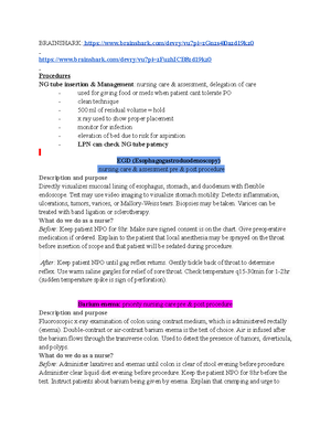
quizlet/517177323/adult-health-1-exam-2-flash-cards/?i=utdiq&x=1jqY Cardiac Testing Cardiac catheterization ● Description & Purpose: Involves insertion of catheter into heart to obtain information about O2 levels and pressure readings within heart chambers. Contrast medium is injected to assist in seeing structures and motion of the heart. Procedure is done by insertion of a catheter into a vein (for right side of heart) and/or an artery (for left side of heart). Preprocedure: -Assess for allergies, especially up contrast dye -Perform baseline assessment, including vital signs, pulse oximetry, heart and breath sounds, neurovascular assessment of extremities (distal pulse, skin temp, skin color, sensation) -Withhold food and fluids for 6-12 hours before.
- assess baseline lab values (cardiac biomarker, creatinine) -Teach patient and caregiver about procedure and post procedure care. Explain the use of local anesthesia at insertion site, placement of catheter, flushed feeling when dye is injected, and possible fluttering sensation of heart as catheter is passed. -Give sedative and other drugs as ordered. Postprocedure: -Perform assessment and compare to baseline: vital signs, pulse oximetry, and heart and breath sounds. Note hypotension or hypertension and signs of pulmonary emboli (respiratory difficulty) -Assess neurovascular status, including peripheral pulses, color, and sensation, of extremity per agency protocol. -Place compression device over arterial site to achieve hemostasis, if indicated. -Observe insertion site for hematoma and bleeding every 15min for the first hour, then according to agency policy. -Monitor ECG for dysrhythmias or other changes (ST segment elevation) -Monitor patient for chest pain and other sources of pain or discomfort. -maintain bedrest as ordered after femoral access. -Maintain IV and/or oral fluid intake and monitor urine output. -Teach patient and caregiver about discharge care, including signs and symptoms to report to HCP(site complications, return of chest pain) and any activity restrictions. -Complications of cardiac catheterization include bleeding or hematoma at the puncture site; allergic reactions to the contrast media; looping or kinking of the catheter; infection; thrombus formation; aortic dissection; dysrhythmias; MI; stroke; and puncture of the ventricles, septum, or lung tissue ● Nursing Management Before: Check for sensitivity to contrast media.. Explain the procedure to the patient including use oWithhold food and fluids for 6-12 hr before the procedure local anesthesia at insertion site, placement of catheter, flushed feeling when dye is injected, and possible fluttering sensation of heart as catheter is passed. Note that a patient may be asked to cough or take a deep breath when dye is injected and that patient is monitored by ECG throughout the procedure. Give sedative and other drugs, as ordered.
After: Frequently assess circulation to extremity used for catheter insertion. Check peripheral pulses, color, and sensation of extremity per agency protocol. Observe
insertion site for hematoma and bleeding. Place compression device over arterial site to achieve hemostasis, if indicated. Monitor vital signs and ECG. Assess for hypotension or hypertension, dysrhythmias, and signs of pulmonary emboli (e., respiratory difficulty).
Stress test
Various protocols are used to evaluate the effect of exercise tolerance on cardiovascular function. A common protocol uses 3-min stages at set speeds and elevation of treadmill belt. The patient can exercise to either predicted peak HR (calculated by subtracting the person's age from 220) or to peak exercise tolerance, at which time the test is ended. The test is also ended for chest discomfort, significant changes in vital signs from baseline, or significant ECG changes (e., ischemia, dysrhythmias). Vital signs and ECG are monitored. The ECG is monitored after exercise for rhythm disturbances or, if ECG changes occurred with exercise, for return to baseline. Continual monitoring of vital signs and ECG for ischemic changes is important in the diagnosis of CAD. An exercise bike may be used if the patient is unable to walk on the treadmill
What should the nurse do? Before : Tell patients to wear comfortable clothes and shoes that can be used for walking or running. Tell the patient about the procedure and importance of reporting any symptoms that occur. β-Blockers may be held 24 hr before the test because they blunt the HR and limit the patient's ability to achieve maximal HR. Caffeine-containing food and fluids are held for 24 hr. Patients must refrain from smoking and strenuous exercise for 3 hr before the test.
During : Monitor vital signs and obtain 12-lead ECG before exercise, during each stage of exercise, and after exercise until all vital signs and ECG changes have returned to normal or baseline. Monitor patient's response throughout the procedure for any signs of distress (e., angina, shortness of breath).
Coronary Artery Bypass Graft (CABG)
● Focus your care on assessing the patient for bleeding (e., chest tube drainage, incision sites), hemodynamic monitoring, checking fluid status, replacing blood and electrolytes as needed, and restoring temperature (e., warming blankets). ● Postoperative dysrhythmias, especially atrial dysrhythmias (e., atrial fibrillation [AF]), are common in the first 3 days after CABG surgery ● This includes impairment of memory, concentration, language comprehension, and social integration. Patients may cry or become teary. Postoperative cognitive dysfunction (POCD) can manifest days to weeks to months after surgery and may become a chronic disorder Discharge instructions
Echocardiogram Ø Ultrasound waves record movement of heart structures; with or without contrast Ø Determines abnormalities of: - Valve structures and motion - Heart chamber size and contents - Ventricular and septal motion and thickness - Pericardial sac - Ascending aorta Ø Measures ejection fraction (EF)—% of end-diastolic blood volume ejected during systole
Lab values
HDL Female:above 55 Male:above 45 ● Lipid profile: Cholesterol, LDL, HDL
● Triglycerides and client education- Normal levels: <150 mg/dL (<1/L) and this will vary with age. Before: Explain procedure to the client and that blood will be sent to the lab. Encourage the client to not drink 24 hours prior to the procedure. During: Blood can be collected in the non fasting stage but varies depending on protocol, some place requires fasting stage. 1. avoid foods high in triglycerides 2. exercise regularly 3. avoid sugar and refined carbs 4. lose weight 5. choose healthier fats like nuts and fish 6. limit alcohol intake 7. eat more vegetables and fruits 8. lifestyle changes
● Normal BUN & creatinine-relationship to hypertension- Hypertension is the leading cause of chronic kidney disease, indications of kidney disease includes an elevated serum creatinine and BUN levels. ●
BUN 8-
Creatine Male- 0-1.
Female- 0-1.
●
● Therapeutic range for INR-relationship to coagulation.0.8-1 for blood disorders is 2- 3. ● PT 11-12 seconds and aPTT 30-40 seconds ● Normal potassium level (3.5-5) ● Normal hemoglobin level
HGB Male- 14-
Female-12-
Hypertension
Etiology:Hypertension can result from either primary or secondary causes.
Primary Hypertension: Primary (essential or idiopathic) hypertension is elevated BP without an identified cause, and it accounts for 90% to 95% of all cases of hypertension.
Organs affected by HYPERTENSION (HIGH BLOOD PRESSURE)
Diagnostic tests
No lab test exists to diagnose hypertension, however several lab tests exist that can ID the cause of secondary hypertension and target organ damage. ● BUN, creatinine- Elevation is indicative of kidney disease ● Elevated blood corticoids- Can indicate cushing’s disease ● Blood glucose and cholesterol studies-ID contributing factors related to blood vessel changes. ● cbc,abg, electrolytes Risk factors 1. age 2. gender 3. family history 4. smoking 5. diabetes 6. dylipidemia
Management
9. Weight loss 10. Limit alcohol use 11. Increase aerobic exercise and activity 12. Reduce sodium intake 13. Maintain adequate sources of dietary potassium, calcium and magnesium 14. Smoking cessation 15. Reduce intake of saturated fats and cholesterol
Heart Failure
Heart Failure - Left side POACHED P pulmonary congestion O rthopnea A dventitious breath sounds C ough H emoptysis E xtreme weakness D yspnea
Differentiate between Right and Left Side Heart Failure (Clinical signs and symptoms)
Differentiate between...
Rheumatic heart disease rheumatic heart disease is a chronic condition resulting from RF (rheumatic fever- acute inflammatory disease of the heart) that is characterized by scarring and deformity of the heart valves affects young adults
Assessment findings : Fever, Subcutaneous nodules and erythema marginatum,Tachycardia, pericardial friction rub, muffled heart sounds, murmurs, peripheral edema,Chorea (involuntary, purposeless, rapid motions; facial grimaces), Signs of monoarthritis or polyarthritis, including swelling, heat, redness, limitation of motion (especially of knees, ankles, elbows, shoulders, wrists)
Nursing management The primary goals of managing a patient with RF are to (1) control and remove the infecting organism; (2) prevent heart complications; and (3) relieve joint pain, fever, and other symptoms. Give antibiotics as ordered to treat the streptococcal infection. Teach the patient that completing the full course of antibiotics is vital to successful treatment. Give salicylates, NSAIDs, and corticosteroids as ordered and monitor fluid intake as appropriate.
Myocarditis
adults or those who are immunocompromised hemorrhages (black longitudinal streaks) that may occur in the nail beds. Petechiae may result from fragmentation and microembolization of vegetative lesions.
Nursing management Tell the patient to avoid people with infections, especially upper respiratory tract infections, and to report cold, flu, and cough symptoms. Stress the importance of avoiding excessive fatigue, and the need to plan rest periods before and after activity. Good oral hygiene, including daily care and regular dental visits, is critical. treatment with antibiotics for 4 to 6 weeks The patient with IE needs adequate periods of physical and emotional rest. Bed rest may be necessary when the patient has fever or complications
Understand cardiac biomarkers: Troponin, CK, and BNP
Cardiac-specific troponin is a heart muscle protein released into circulation after injury or infarction. Normally the level in the blood is very low, so a rise in level is diagnostic of myocardial infarction (MI) or injury. cTnT and cTnI are detectable within hours (on average 4 to 6 hours) of MI or injury, peak at 10 to 24 hours, and can be detected for up to 10 to 14 days.
Troponin is the biomarker of choice in the diagnosis of ACS. High-sensitivity troponin (hs- cTnT, hs-cTnI) assays may provide even earlier detection of a heart event. The normal range for troponin is between 0 and 0 ng/mL. CTNI negative(<0,5) Indeterminate or suspicious for injury to myocardium: (0.5-2) Positive for myocardial injury (>2) CTNT: <0.
Creatine kinase (CK) enzymes are found in a variety of organs and tissues and occur as three isoenzymes. These isoenzymes are specific to skeletal muscle (CK-MM), brain and nervous tissue (CK-BB), and the heart (CK-MB). CK-MB rise is specific for MI or injury. Levels begin to increase 3 to 6 hours after symptom onset, peak in 12 to 24 hours, and return to baseline within 12 to 48 hours after MI. The peak level and return to normal can be delayed in a patient with a large MI. Levels drop more rapidly in patients who are quickly and successfully treated for an MI.
The natriuretic peptides (atrial natriuretic peptide [ANP] and b-type natriuretic peptide [BNP]) are secreted by heart cells. They antagonize the effects of antidiuretic hormone (ADH) and aldosterone. This results in natriuresis (excretion of sodium in urine) and diuresis, resulting in reduced blood volume and BP.
Pacemaker indications
Understand the treatment for various heart rhythms
V-fib - Treatment consists of immediate initiation of CPR and advanced cardiovascular life support (ACLS) with the use of defibrillation and definitive drug therapy (e., epinephrine, vasopressin). There should be no delay in starting chest compressions and using a defibrillator once available.
V-tach - Precipitating causes (e., electrolyte imbalances, ischemia) must be identified and treated. If the VT is monomorphic (uniform and over 100 bpm) and the patient is clinically stable (i., pulse is present) and has preserved left ventricular function, IV procainamide (antiarrhythmic), sotalol (antiarrhythmic), or amiodarone (antiarrhythmic) is used. These drugs can also be used if the VT is polymorphic with a normal baseline QT interval
Bradycardia - For the patient with symptoms, treatment consists of giving IV atropine (anticholinergic drug). If atropine is ineffective, transcutaneous pacing or a dopamine or epinephrine (Adrenalin) infusion is considered. Permanent pacemaker therapy may be needed. If bradycardia is due to drugs, these may have to be held, discontinued, or reduced.
Tachycardia -The underlying cause of tachycardia guides the treatment. For example, if the patient is experiencing tachycardia from pain, effective pain management is important to treat the tachycardia. In clinically stable patients, vagal maneuvers can be attempted. In addition, IV β- blockers (e., metoprolol [Lopressor]), adenosine (Adenocard), or calcium channel blockers (e., diltiazem [Cardizem]) can be given to reduce HR and decrease myocardial O 2 consumption. In clinically unstable patients, synchronized cardioversion is used.
Coronary Artery Disease
Assessment, and clinical manifestations What is the Plan of Care for this disorder? Client education to minimize complications
Risk factors
Increasing age
Gender (more common in men than in women until 75 yr of age)
Ethnicity (more common in white men than in African Americans)
Genetic predisposition and family history of heart disease
Atherosclerosis
o Begin as soft deposits of fat that harden with age
o Referred to as hardening of the arteries
o Atheroma’s (fatty deposits prefer coronary arteries
- Atherosclerosis is major cause of CAD
o Characterized by lipid deposits within
o HDL greater than 40
o Triglycerides: less than 150
UA
Creatinine
BUN
Electrolytes
Bilateral BP management
ECG
Echocardiogram
Unstable Angina Vs Stable Angina & Explain STEMI vs NSTEMI
A STEMI (the worst) caused by an occlusive thrombus creates ST-elevation in the ECG leads facing the area of infarction
NSTEMI, caused by a partialocclusive thrombus, does not cause ST segment elevation on the 12-lead ECG. Patients may or may not develop ST-T wave changes in the leads affected by the infarction
Stable angina Assessment, and clinical manifestations
Most angina pain occurs substernally, it may radiate to other locations, including the jaw, neck, shoulders, and/or arms. Many people with angina complain of indigestion or a burning sensation in the epigastric region. The sensation may also be felt between the shoulder blades. Often people who complain of pain between the shoulder blades or indigestion type pain dismiss it as not being heart related
- Episodic pain lasting a few minutes
- Provoked by exertion or stress
- Relieved by rest or nitroglycerin
What is the Plan of Care for this disorder?
Antiplatelet/anticoagulant therapy Antianginal therapy ACE inhibitor/angiotensin receptor blocker B β-blocker BP control Cigarette smoking cessation Cholesterol (lipid) management Calcium channel blockers Cardiac rehabilitation Diet (weight management) Diabetes management Depression screening Education Exercise Flu vaccination
Client education to minimize complications Educate on ● Hypertension control ● Reduce fat intake and caloric intake ● Smoking cessation ● increase physical activity ● Take prescribed drugs for diabetes
Unstable angina Assessment and clinical manifestations
- New-onset angina • Chronic stable angina that increases in frequency, duration, or severity • Occurs at rest or with minimal exertion • Lasts more than 10 min
What is the plan of care for this disorder? Acute drug therapy - nitroglycerin - Antiplatelet therapy - Anticoagulation therapy Coronary angiography
Adult health-EXAM 2 -2
Course: Adult Health I (NR-324)
University: Chamberlain University
This is a preview
Access to all documents
Get Unlimited Downloads
Improve your grades
