- Information
- AI Chat
Was this document helpful?
Chapter 7 Cell Strucutre and Function
Course: College Geometry (MAT 115)
4 Documents
Students shared 4 documents in this course
University: Rowan College of South Jersey
Was this document helpful?

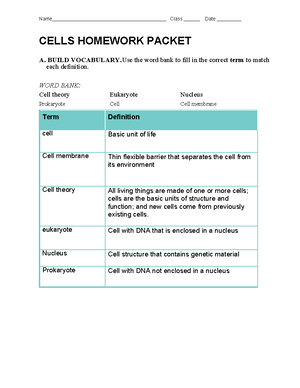
Cell: basic unit of all forms of life
Cell theory: fundamental concept of biology
that states that all living things are composed
of cells; that cells are the basic units of
structure and function in living things; and
that new cells are produced from existing
cells.
Cell membrane: thin, flexible barrier that
surrounds all cells; regulates what enters and
leaves the cell
Nucleus: the center of an atom, which
contains the protons and neutrons; in cells,
structure that contains the cell's genetic
material in the form of DNA.
Eukaryotic: organism whose cells contain a
nucleus
Prokaryote: unicellular organism that lacks
a nucleus
Vocabulary:
Key Questions
All living things are made up of cells.
○
Cells are the basic units of structure and function in living things.
○
New cells are produced from existing cells.
○
The cell theory states: •
What is the cell theory?
Most microscopes use lenses to magnify the image of an object by focusing light or
electrons.
•
How do microscopes work?
Prokaryotic cells do not separate their genetic material within a nucleus.
•
In eukaryotic cells, the nucleus separates the genetic material from the rest of the cell.
•
How are prokaryotic and eukaryotic cells different?
The smallest unit of any organism- the cell.
The Discovery of the Cell
Without the instruments to make them visible, cells remained out of sight and, therefore, out of mind for the most of human history. All of this
changed with a dramatic advance in technology- the invention of the microscope.
Early Microscopes
In 1665, Englishman Robert Hooke used an early compound microscope to look at a nonliving thin slice of cork, a plant material. Under the
microscope, cork seemed to be made of thousands of tiny empty chambers. Hooke called these chambers "cells" because they reminded him of
a monastery's tiny rooms, which were called cells. The term cell is used in biology to this day. Today we know that living cells are not empty
chambers, that in fact they contain a huge array or working parts, each with its own function, (organelle.)
In Holland around the same time, Anton van Leeuwenhoek, used a single lens microscope to observe pond water and other things. To his
amazement, the microscope revealed a fantastic world of tiny living organisms that seemed to be everywhere, in the water he and his
neighbors drank, and even in his own mouth. Leeuwenhoek's illustrations of the organisms he found in the human mouth-- which today we call
bacteria.
Cork under microscope: first "Cells"
Robert Hooke:
Robert Hooke's Microscope
Robert Hooke
Anton van Leeuwenhoek:
Leeuwenhoek's Microscope
7.1 Life is Cellular
Sunday, December 16, 2012
1:07 PM
Chapter 7 Cell Strucutre and Function Page 1
More from:College Geometry(MAT 115)
More from: